

Papilio5D : Highly Sensitive Light Sheet Microscope for Organoids and 3D Cell Cultures
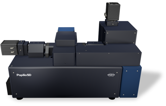
The Bruker Papilio5D light-sheet microscope is engineered to deliver high-fidelity volumetric imaging with exceptional depth penetration across a broad range of biological specimens, including highly scattering organoids and zebrafish larvae. The platform integrates dual-view detection with modular illumination architectures to maximize photon efficiency. Dual-sided detection captures approximately twice the photon yield of single-objective configurations, providing more complete three-dimensional sampling and improved reconstruction accuracy. Users may select Gaussian or Bessel beam illumination, each offering sub-micrometer beam widths. Flexible sample-mounting solutions further support multiplexed experimental workflows and pharmacological perturbation studies.
High-numerical-aperture, water-dipping objectives generate ultra-thin light sheets, while both area- and line-mode detection accommodate diverse sample constraints.
These capabilities collectively enhance signal-to-noise ratios, axial resolution, and imaging depth. The system can achieve Nyquist sampling, enabling advanced deconvolution and high-precision downstream image analysis.
The Papilio5D sample stage accommodates up to three dishes, each subdivided into two independently addressable chambers, allowing six parallel experimental conditions. Long-term specimen viability is supported via comprehensive environmental regulation, with precise control of temperature (20-40 °C), CO₂/O₂ levels, and humidity.
In area-mode acquisition, the camera records the full field of view per exposure, providing high throughput and efficient data capture. In line-mode acquisition, rolling-shutter readout is synchronized with the scanned light sheet to produce a confocal-slit effect that rejects out-of-focus and scattered photons. This mode substantially increases image contrast in thick or highly scattering preparations such as organoids.